Opal-like Structures and Inverse Opal-like Structures
Introduction
Photonic crystals (PCs) with three-dimensional (3D) periodicity of dielectric structures, called artificial opals, or Opal-like Structures, have attracted much attention in the past decade for their ability in light manipulation, which has potential applications in the area of photonics [1]. The inverse opal-like structures (inverse OLS) can be used as eliminators of unwanted heat from the thermal emission sources [2], in the conventional lighting field for increasing efficiency and obtaining improved thermophotovoltaic devices [3], as piezoelectric transducers, solar cells, phosphors, short-wavelength light emitters [4], gas sensors [5] and so on.
Fabrication
Highly ordered artificial opals can be fabricated by using high quality monodisperse spheres made of silica, polymethylmethacrylate, or polystyrene with a procedure based on combination of self-assembly and sol–gel method (sedimentation) [1, 6, 7] (Fig. 1a), self-assembly and pressure (colloidal epitaxy) [8-10] (Fig.1b), self-assembly onto polished conductive substrates by the vertical deposition method (convective assembly, or controlled drying) [11-16] (Fig.1c), spin coating [17], electrophoresis [18-20].
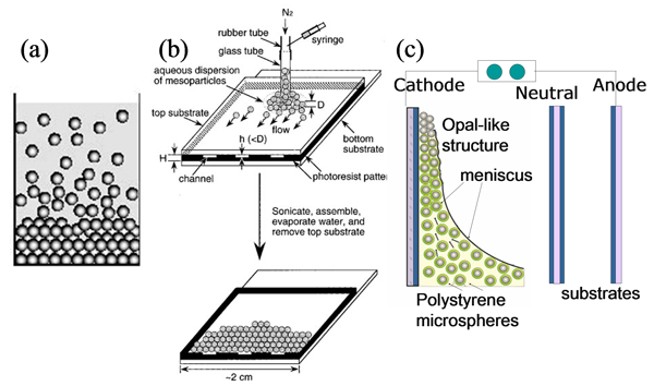
Figure 1. Methods of fabrication the opal-like structures
If the voids of artificial opals used as templates are filled with other materials – polymers [21-23], liquids [24-26], inorganic oxides [27-30], carbon [31], semiconductors [32-35], superconductors [36-38], and metals [39-45] and then the templates is removed (if necessary) we have got an inverse opal-like structures.
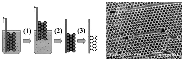
Figure 2. Fabrication (1-3) and SEM image of the inverse OLS.
On Figure 3 the features of the natural and artificial opals are presented.
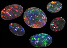
Figure 3. The features of the natural (a) and artificial (b) opals.
Structure
The scanning electron microscopy (SEM) image of the synthesized samples is presented in Fig.1. Typically the vertical deposition method produces a thin OLS consisting of 10 – 40 layers on the substrate. The geometry of the synthesis directly determines the geometry of the future crystal. The direction perpendicular to the substrate is the always the [111] axis of the FCC structure. The vertical axis (axis of aqueous suspension drying, or meniscus moving) always corresponds to the [22-2] crystallographic direction.
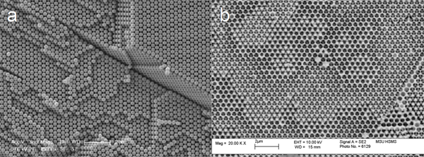
Figure 4. SEM images of the top view of OLS (a) and inverse OLS on the base of Co (b) films.
Thus, already on the stage of synthesis one is able to have a clear understanding of the orientation of the crystal. The question remained unsolved is about the structural quality of the obtained crystals which can degrade at each step of the synthesis: 1) synthesis of the template, 2) synthesis of inverse structure, 3) dissolvent of the template.
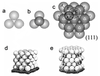
Figure 5. Forming of the hard package structure of the microsphere: (a) first level (A), (b) second level (B) and (c) third level (C); (d) FCC structure; (e) HCP structure.
The silica or polystyrene spheres typically pack in a face-centered cubic structure, wherein each sphere contacts 12 others (6 in the same layer, 3 in the layer above and 3 in the layer below) (Fig. 5). Hence, the voids of the artificial opal have quasi-cubic and quasi-tetrahedral forms with concave sides and they are connected by vertices along each of four [111] axes (Fig. 6). It means, that we can considered inverse OLS as an assembly of small metallic particles duplicating the shape of the voids and connected to each other via thin (several tens of nanometers) and long (several hundreds of nanometers) “legs”.
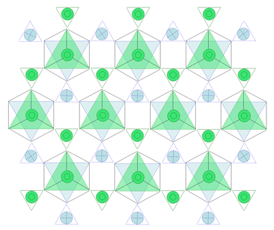
Figure 6. Schema of the quasi-cubic and quasi-tetrahedral voids of OLS connected by vertices along one of [111] axes.
Studying the internal structure of photonic crystals is inherently difficult: their optical properties exclude the use of microscopy or light scattering techniques for detailed characterization. A similar argument holds for electron microscopy techniques. Using these, information can only be gained about the surface structure of the crystals. X-rays, however, do have the required penetration depth to study the interior of these crystals. By using a specialized X-ray diffraction setup at BM-26 DUBBLE (European Synchrotron Radiation Facility, Grenoble, France) [46, 47] , the internal structure of OLS and inverted crystals was probed. Since the particles are larger than 400 nm in diameter, an extremely high resolution in reciprocal space is required in order to be able to study the crystals using radiation with a wavelength of the order of one Ångström. This is achieved by focusing the beam at the detector using a set of Beryllium compound refractive lenses positioned next to the sample [48, 49].
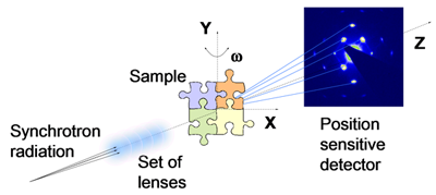
Fig. 7. Schematic drawing of the diffraction experiments with the use of synchrotron radiation.
The diffraction patterns are recorded at various sample rotation angles of -65o < w < +65° around the [20-2] crystallographic direction, where w = 0° corresponds to the geometry of the film surface perpendicular to the beam (Fig. 7). It is allowed us to obtain the information about ordering the samples in different crystallographic directions.
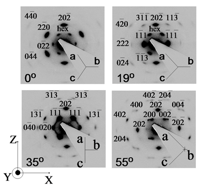
Fig. 8. The diffraction patterns of the OLS at rotation angles w = 0°(a), 19°(b), 35°(c), and 55°(d).
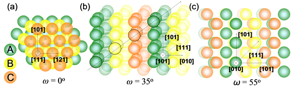
Fig. 9. Real space OLS orientations
The surprise is to be found in Figure 8c, measured along the [101] crystallographic direction of FCC. Most peaks do indeed correspond to this, but in addition there are diffuse scattering stripes in the images between 111 and 020 types of reflections. These are the so-called Bragg rods and are indicative of stacking disorder. It is well seen form Figure 9b that the small moving of the colloidal spheres banded by black line from (½,½,1) to (½, ½, ½) positions (along [00-1] direction on Fig.9b) will be result into stacking faults along [111] direction. These findings showed that the line defects, which are usually seen at the crystal surface with scanning electron microscopy (Figure 10), in fact correspond to two-dimensional stacking fault defects terminating at the crystal surface.
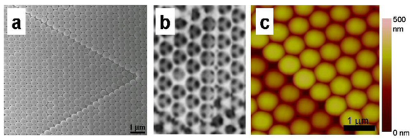
Fig. 10. Real space crystal structures. Panel (a) and (b) show SEM pictures of an as-grown crystal and an inverted crystal respectively. The lines of square packing correspond to the stacking faults. In the inverted crystal (b), the same square configuration can be observed in the second layer. Panel (c) is an AFM image, showing the height step associated with the stacking faults.
Except one- and two-dimensional stacking fault defects there are numbers of faults of different nature such as cracks, dislocations, twin planes, point defects. The photonic crystal engineering can use a number of parameters, which can influence the forces involved in the colloid self-assembly process and responsible for the formation of OLS. For example, by adjusting the density mismatch between the particles and the solvent, one can modify the strength of the effective gravity force. Variation of the growth temperature can slightly affect the diffusion and can have a strong effect on the evaporation rate and, therefore, on the convection force. Changing the particle charge can influence the interparticle forces. Still, one may need to have additional means to vary the force balance and, therefore, to fine tune the crystal structure and quality.
Electric field could be an excellent tool to control the crystallization process. Here we present results on application of DC electric field normal to the substrates during vertical deposition of colloidal crystals (Fig.1c). Despite the negative charge of the particles, the crystals are found to form on both positively- and negatively-charged electrodes. It is found that electrostatic attraction between the particles and the substrate increases the crystal thickness while repulsion between them promotes higher crystal quality.
The representative scanning electron micrographs of as-grown colloidal crystals of polystyrene spheres obtained on a cathode and on an anode are shown in Fig. 11. Panels A and B correspond to the top surface and cross section of the crystal deposited at U = 1.5 V on a cathode, C and D – at 3 V on an anode. On the cathode one can see that the particles are neatly arranged into periodic arrays.
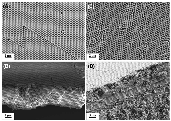
Fig.11. SEM images of OLS prepared by vertical deposition method in a presence of an external electric field perpendicular to the substrates. Panels A and B correspond to the top surface and cross section of the crystal deposited at U = - 1.5 V, C and D – at U = +3 V.
Still, some defects are visible such as a few vacancies and two-dimensional stacking fault (lines) (Fig. 11a). Fig. 11c illustrates that anode polarization leads to formation of highly defective colloidal crystal structure. In this case the electrostatic attraction between negatively charged particles and the positive electrode is presumably too strong so that the polystyrene spheres have little chance to rearrange their position to find a better place in the crystal. Only top layers of the colloidal crystals formed on are ordered due, probably, to screening of Coulomb interaction by the bottom layers (Fig. 11d).
The results of the microradian X-ray diffraction are shown in Fig.12. In the diffraction patterns one can clearly identify a large number of Bragg reflections, which can be assigned to the reciprocal lattice of the ideal FCC crystal structure with the cubic cell size of a0 = 750 nm. It is seen, as well, that diffraction pattern for OLS synthesized at U = -1.5V consists of pointed high order reflections up to four order in compared to artificial opal synthesized at U = +1.5V.
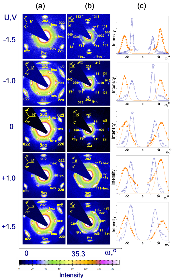
Fig. 12. The diffraction patterns of the OLS at rotation angles w = 0° (a) and 35° (b) for samples synthesized at different U = -1.5, -1, 0, +1, and +1.5 V. (c) The w-dependence of the intensity for (-11-1), and (-1-11) Bragg reflections.
The monotonic improvement of the crystal quality from U = +1.5.V to U = -1.5 V can also be derived from the width of the diffraction spots (Fig.12c). The results obtained for 111 reflection are summarized in Fig. 13b. The full-width at half-maximum (FWHM) of the diffraction maxima in the azimuthal (Δq) and radial (δq) directions characterizes the mosaicity of the colloidal films and the average crystallite size (Λ), respectively. One can see that the mosaicity Δq of OLS decreases from 8º to 4º when the applied voltage changes from +1.5 V to -1.5 V. The peak width in the radial direction is determined from the fit by a Lorenzian accounting for the instrument resolution value (FWHM of the profile of the direct beam). One can see in Fig. 13b that application of the negative potential leads to a significant increase of the average size of crystallites Λ = 2πB/δq, where B is a factor of the order 1.
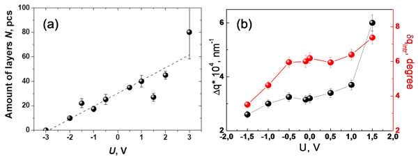
Fig. 13. a) The dependence of thickness of a colloidal film on applied voltage U (according to SEM data Fig.13). b) The longitudinal and transfers width of the reflection (111) on the potential applied upon OLS synthesis.
Thus, increase of a cathode polarization leads to the improvement of crystal quality but, at the same time, the thickness of a film decreases (see Fig. 11b,d). Optimum value of an applied voltage U for the conditions we have used (given concentration of the suspension, pH, charge of colloidal particles, temperature, distance between electrodes, etc.) is around -1.5 V. These conditions allow growing crystals that are 20 layers thick.
Magnetic Structure of OLS on the based of ferromagnetic materials
The detailed picture of the transformation of the magnetic structure under in-plane applied field was detected with help of small-angle diffraction of polarized neutrons (polarized SANS). SANS measurements were carried out with the instrument SANS-2 at the Geesthacht Neutron Facility (GeNF) [50]. The schematic drawing of the setup is shown in Fig.14. It points out that the instrument equipped with the Polarizer and the Spin Flipper to provide the possibility studying the polarization dependent neutron cross section.
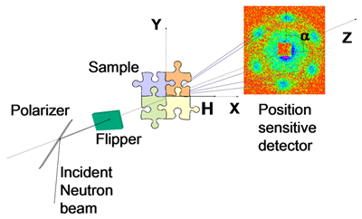
Fig. 14. Schematic drawing of the polarized neutron diffraction experiment.
In our experiments the neutron scattering intensities I+(q,+P0) and I–(q,-P0) were measured with the neutron beam polarized parallel and antiparallel to the external magnetic field, respectively. An external magnetic field was applied perpendicular to incident beam and ranged from -200 mT to +200 mT. The cross section of polarized neutrons from a magnetic structure with a large period can be split into three contributions [51]:
nuclear scattering
I(q) = ½(I+(q,+P0) + I-(q,-P0)) ~ |AnS(q)F(q)|2 + |AmmqS(q)F(q)|2. (1)
represents the sum of the nuclear and magnetic scattering (independent of neutron polarization P);
magnetic scattering
Im(q)=I(q,m(H))–I(q,m(Hc))~ |AmmqS(q)F(q)|2 (2)
is proportional to the difference between the magnetic cross-sections of the sample in two principally different states: partially magnetized in the finite field H and fully demagnetized at H ~Hc (independent of neutron polarization P);
interference scattering
dI(q) = I+(q,+P0) - I-(q,-P0) ~ 2(P0<m>q)AnAm|S(q)F(q)|2 (3).
It is attributed to the nuclear-magnetic interference indicating the correlation between the magnetic and nuclear structures (dependent of neutron polarization P).
The neutron diffraction patterns are shown in Fig.15. Observed reflections correspond to the neutron scattering on the {202} crystallographic planes what is in a good agreement with microradian X-ray diffraction data (Fig.8 a). The external magnetic field was applied in two different geometries: H along the [-12-1] axis (geometry I, Fig.15 a) and H along the [-110] axis (geometry II, Fig.15 b) to investigate influence of the structural anisotropy on behavior of the magnetization vector in the reversal magnetization process. In order to appropriately estimate the magnetic contribution Im(q) and interference contribution DI(q) into the scattering, one has to perform the analysis of these intensities as a function of the azimuth angle a, where a is the angle between the direction of the scattering vector q and the field direction H, which points along the horizon in the detector plane (Fig. 15).
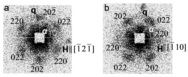
Fig. 15. The neutron scattering map for inverse Co OLS sample for (a) geometry I and (b) geometry II at H = 200 mT.
The more detailed information will be present on this site after publication of these materials in the article N.A. Grigoryeva, A.A. Mistonov, K.S. Napolskii, N.A. Sapoletova, A.A. Eliseev, A.V. Vasilieva, A.V. Petukhov, D. Byelov, D.Yu. Chernyshov, H. Eckerlebe, S.V. Grigoriev, “Magnetic topology of Co based inverse opal-like structures” Phys. Rev. B, (2010). In general, the complex magnetic structure of the OLS on the based of ferromagnetic materials can be interpreted in terms of non-homogeneous distribution of magnetization density within the unit element of opal-like structure. As was mentioned above, the unit element consists of three parts: quasi-tetrahedral, quasi-cubic and quasi-tetrahedral which are connected by vertices along the [111] axes. Such non-homogeneous distribution is determined by the combined effect of the easy-plane geometry of the sample film itself and the crystallographic geometry of the opal-like structure with respect to the magnetic field axis.
References.
1. Advanced Materials, 13, №6 ,369 – 450. Photonic crystals are the central theme of this issue.
2. J.G. Fleming, S.Y. Lin I. El-Kady, R. Biswas, K.M. Ho, Nature, 2002, 417, 52.
3. S.Y. Lin, J. Moreno, J.G. Fleming, Appl. Phys. Lett. 2003, 83, 380.
4. D. C. Look, Mater. Sci. Eng. B, 2001, 80, 383.
5. C. Baratto, G. Faglia, G. Sberveglieri, A. Sutti, G. Calestani, C. Dionigi, IEEE Sensors, 2005, 1196 – 1200.
6. Waterhouse, G. I. N.; Waterland, M. R. Polyhedron 2007, 26, 356-368.
7. Miguez, H.; Lopez, C.; Meseguer, F.; Blanco, A.; Vazquez, L.; Mayoral, R.; Ocana, M.; Fornes, V.; Mifsud, A. Appl. Phys. Lett. 1997, 71, 1148-1150.
8. Sang Hyun Park and Younan Xia, Langmuir 1999, 15, 266-273
9. Hoogenboom, J. P.; van Langen-Suurling, A. K.; Romijn, J.; van Blaaderen, A. Phys. Rev. E 2004, 69, 051602.
10. van Blaaderen, A.; Ruel, R.; Wiltzius, P. Nature 1997, 385, 321-324.
11. K.S. Napolskii, N.A. Sapoletova, D.F. Gorozhankin, A.A. Eliseev, D.Yu. Chernyshov, D.V. Byelov, N.A. Grigoryeva, A.A. Mistonov, W.G. Bouwman, K.O. Kvashnina, A.V. Lukashin, A.A. Snigirev, A.V. Vassilieva, S.V. Grigoriev, AV. Petukhov. Fabrication of Artificial Opals by Electric-Field-Assisted Vertical Deposition // Langmuir, 2009, DOI: 10.1021/la902793b
12. Jiang, P.; Bertone, J. F.; Hwang, K. S.; Colvin, V. L. Chem. Mater. 1999, 11, 2132-2140.
13. Blanco, A.; Chomski, E.; Grabtchak, S.; Ibisate, M.; John, S.; Leonard, S. W.; Lopez, C.; Meseguer, F.; Miguez, H.; Mondia, J. P.; Ozin, G. A.; Toader, O.; van Driel, H. M. Nature 2000, 405, 437-440.
14. Norris, D. J.; Arlinghaus, E. G.; Meng, L.; Heiny, R.; Scriven, L. E. Adv. Mater. 2004, 16, 1393-1399.
15. Meng, L.; Wei, H.; Nagel, A.; Wiley, B. J.; Scriven, L. E.; Norris, D. J. Nano Lett. 2006, 6, 2249-2253.
16. Wei, H.; Meng, L.; Jun, Y.; Norris, D. J. Appl. Phys. Lett. 2006, 89, 241913.
17. Jiang, P.; McFarland, M. J. J. Am. Chem. Soc. 2004, 126, 13778-13786.
18. Trau, M.; Saville, D. A.; Aksay, I. A. Langmuir1997, 13, 6375-6381.
19. Rogach, A. L.; Kotov, N. A.; Koktysh, D. S.; Ostrander, J. W.; Ragoisha, G. A. Chem. Mater. 2000, 12, 2721-2726.
20. Holgado, M.; Garcia-Santamaria, F.; Blanco, A.; Ibisate, M.; Cintas, A.; Miguez, H.; Serna, C. J.; Molpeceres, C.; Requena, J.; Mifsud, A.; Meseguer, F.; Lopez, C. Langmuir 1999, 15, 4701-4704.
21. S.A. Jonson, P.J. Ollivier, T.E. Mallouk, Science 1999, 283, 963.
22. P. Jiang, K.S. Hwang, D.M. Mittleman, J.E. Bertone, V.L. Colvin, J. Am. Chem. Soc. 1999, 121, 11630-11637.
23. T. Simuda, Y. Wada, T. Kitamura, S. Yanagida, Chem. Commun. 2000, 1613.
24. K. Busch, S. John, Phys. Rev. Lett. 1999, 83, 967-970.
25. E. Yablonovitch, Nature, 1999, 401, 539-541.
26. O. K. Alimov, T. T. Basiev, Yu. V. Orlovskii, V. V. Osiko, M. I. Samoilovich, Quantum Electron. 2008, 38, 665-669.
27. B.T. Holland, C.F. Blanford, A. Stein, Science 1998, 281, 538.
28. J.E.G.J. Wijnhoven, W.L. Vos, Science 1998, 281, 802.
29. H. Yan, C.F. Blanford, B.T. Holland, W.H. Smyrl, A. Stein, Chem. Mater. 2000, 12, 1134.
30. F. Chen, C. Xia, M. Liu, Chem. Lett. 2001, 1032.
31. A.A. Zakhidov, R.H. Baughman, Z. Iqbal, C. Cui, I. Khayrullin, S.O. Dantas, J. Marti, V.G. Ralchenko, Science 1998, 282, 897.
32. Y.A. Vlasov, N. Yao, D.J. Norris, Adv. Mater. 1999, 11, 165.
33. P.V. Braun, P. Wiltzius, Nature, 1999, 402, 603.
34. A. Blanco, E. Chomski, S. Grabtchak, M. Ibisate, S. John, S.W. Leonard, C. Lopez, F. Meseguer, H. Miguez, J.P. Mondia, G.A. Ozin, O. Toader, H.M. van Driel, Nature, 2000, 405, 437.
35. Z. Lei, J. Li, Y. Ke, Y. Zhang, H. Wang, G. He, J. Mater. Chem. 2001, 11, 1778.
36. A. Lungu, M. Bleiweiss, J. Amirzadeh, S. Saygi, A. Dimofte, M. Yin, Z. Iqbal, T. Datta. Physica C, 2001, 349, 1.
37. A.A. Zhukov, E.T. Filby, M.A. Ghanem, P.N. Barlett, P.A.J. de Groot, Physica C, 2004, 404, 455.
38. A.E. Aliev, S.B. Lee, A.A. Zakhidov, R.H. Baughman, Physica C, 2007, 453, 15.
39. O.D. Velev, P.M. Tessier, A.M. Lenhoff, E.W. Kaner, Nature, 1999, 401, 548.
40. P. Jiang, J. Cizeron, J.F. Bertone, V.L. Colvin, J. Am. Chem. Soc. 1999, 121, 7957.
41. H. Yan, C.F. Blanford, B.T. Holland, M. Parent, W.H. Smyrl, A. Stein, Adv. Mater. 1999, 11, 1003.
42. L. Xu, W.L. Zhou, C. Fromer, R.H. Baughman, A.A. Zakhidov, L. Malkinski, J.-Q. Wang, J.B. Wiley, Chem. Commun. 2000, 997.
43. J.E.G.J. Wijnhoven, S.J.M. Zevenhuizen, M.A. Hendriks, D. Vanmaekelbergh, J.J. Kelly, W.L. Vos, Adv. Mater. 2000, 12, 888.
44. P.N. Bartlett, P.R. Birkin, M.A. ghanem, Chem. Commun. 2000, 1671.
45. L. Xu, L. D. Tung, L. Spinu, A.A. Zakhidov, R.H. Baughman, J.B. Wiley, Adv. Mater. 2003, 15, №18, 1562-1564.
46. DUBBLE Beamline BM26: http://www.esrf.eu/UsersAndScience/Experiments/CRG/BM26
47. J. H. J. Thijssen, A. V. Petukhov, D. C. ’t Hart, A. Imhof, C. H. M. Van der Werf, R. E. I. Schropp, A. Van Blaaderen, Adv. Mater., 18, 1662 (2006)
48. A.V. Petukhov, D.G.A.L. Aarts, I.P. Dolbnya, E.H.A. de Hoog, K. Kassapidou, G.J. Vroege, W. Bras, H.N.W. Lekkerkerker, Phys.Rev.Lett. 88, 208301 (2002).
49. A.V. Petukhov, I.P. Dolbnya, D.G.A.L. Aarts, G.J. Vroege, H.N.W. Lekkerkerker, Phys.Rev.Lett. 90, 028304 (2003).
50. http://www.gkss.de/pages.php?page=w_abt_genesys_sans-2.html&language=d&version=g
51. S.V. Grigoriev, K.S. Napolskii, N.A. Grigoryeva, A.V. Vasilieva, A.A. Mistonov, D.Yu. Chernyshov, A.V. Petukhov, D.V. Byelov, A.A. Eliseev, A.V. Lukashin, Yu.D. Tretyakov, A.S. Sinitskii, and H. Eckerlebe, Phys. Rev. B, 79, 045123(8) (2009).
The results of the investigation artificial opals and inverse opal-like structures are published in:
1. Kirill S. Napolskii, Nina A. Sapoletova, Dmitriy F. Gorozhankin, Andrey A. Eliseev, Andrei V. Petukhov, Dmytro V. Byelov, Alexander A. Mistonov, Natalia A. Grigoryeva, Wim G. Bouwman, Kristina O. Kvashnina, Anatoly A. Snigirev, Dmitry Yu. Chernyshov, Alexandra V. Vassilieva, Sergey V. Grigoriev, Fabrication of Artificial Opals by Electric-Field-Assisted Vertical Deposition, Langmuir, 2010, v. 26(4), pp. 2346-2351.
2. S. V. Grigoriev, K. S. Napolskii, N. A. Grigoryeva, A. V. Vasilieva, A. A. Mistonov, D. Yu. Chernyshov, A. V. Petukhov, D. V. Belov, A. A. Eliseev, A. V. Lukashin, Yu. D. Tretyakov, A. S. Sinitskii, and H. Eckerlebe, “Study of inverse Ni-based photonic crystal using the microradian X-ray diffraction”, J. of Phys.: Confer.Ser., v.247, 012029 - 012035 (2010).
3. Саполетова Н.А., Маргынова Н.А., Напольский К.C., Елисеев А.А., Лукашин А.В., Петухов Д.И., Кушнир С.Е., Васильева А.В., Григорьев С.В., Н.А. Григорьева, Мистонов А.A., Белов Д.В., Третьяков Ю.Д., Самосборка коллоидных частиц в присутствии электрического поля, ФТТ, 2011, т. 53, вып.6, 1064-1068.
4. N.A. Grigoryeva, A.A. Mistonov, K.S. Napolskii, N.A. Sapoletova, A.A. Eliseev, A.V. Vasilieva, W. Bouwman, D.V. Byelov, A.V. Petukhov, D. Yu. Chernyshov, H. Eckerlebe, S.V. Grigoriev «Magnetic topology of Co based inverse opal-like structure», Physical Review B, 84, 2011, 064405(13).
5. K.S. Napolskii, I.V. RoslyakovA.A. Eliseev, D. Byelov, , A.V. Petukhov, N.A. Grigoryeva, W.G. Bouwman, A.V. Lukashin A.P. Chumakov, and S.V. Grigoriev, "The Kinetics and Mechanism of Long-Range Pore Ordering in Anodic Films on Aluminum", The Journal of Physical Chemistry, 2011.
6. А.В. Васильева, С.В. Григорьев, Н.А. Григорьева, А.А.Мистонов, К.С.Напольский, Н.А.Саполетова, А.В. Петухов, Д.В.Белов, А.А. Елисеев, Д.Ю.Чернышов, А.И.Окороков, Анализ дефектности опалоподобных фотонных кристаллов, синтезированных на проводящих подложках, Физика твердого тела, 2010, том 52, вып. 5, c. 1017-1020
7. A. Sinitskii, V. Abramova, N. Grigorieva, S. Grigoriev, A., Snigirev, D. V. Byelov and A. V. Petukhov, Revealing stacking sequences in inverse opals by microradian X-ray diffraction, EPL, 89 (2010) 14002.
8. В.В. Абрамова, А.С. Синицкий, Н. А. Григорьева, С. В. Григорьев, Д. В. Белов, А. В. Петухов, А. А. Мистонов, А. В. Васильева, Ю. Д. Третьяков, Анализ структуры фотонных кристаллов методом ультрамалоуглового рентгеновского рассеяния, ЖЭТФ, 2009, том 136, вып.1(7), стр.1‑7. English version: V. V. Abramova, A. S. Sinitskii, N. A. Grigor’eva, S. V. Grigor’ev, D. V. Belov, A. V. Petukhov, A. A. Mistonov, A. V. Vasil’eva, and Yu. D. Tret’yakov, «Ultrasmall_Angle X_ray Scattering Analysis of Photonic Crystal Structure» Journal of Experimental and Theoretical Physics, 2009, Vol. 109, No. 1, pp. 29–34.
9. А.А.Елисеев, Д.Ф. Горожанкин, К.С. Напольский, А.В. Петухов, Н.А. аполетова, А.В. Васильева, Н.А. Григорьева, А.А. Мистонов, Д.В. Белов, В.Г. Бауман, К.О. Квашнина, Д.Ю. Чернышов, А.А. Босак, С.В. Григорьев, «Определение реальной структуры искусственных и природных опалов на основе трехмерных реконструкций обратного пространства», 2009, Письма в ЖЭТФ, т.90, вып.4, с.297-303.
10. Jan Hilhorst, Vera V. Abramova, Alexander Sinitskii, Nina A. Sapoletova, Kirill S. Napolskii, Andrey A. Eliseev, Dmytro V. Byelov, Natali A. Grigoryeva, Alexandra V. Vasilieva, Wim G. Bouwman, Kristina Kvashnina, Anatoly Snigirev, Sergey V. Grigoriev, Andrei V. Petukhov†, «Double Stacking Faults in Convectively Assembled Crystals of Colloidal Spheres», Langmuir 2009, 25(17), 10408–10412
11. S. V. Grigoriev, K. S. Napolskii, N. A. Grigoryeva, A. V. Vasilieva, A. A. Mistonov, D. Yu. Chernyshov, A. V. Petukhov, D. V. Belov, A. A. Eliseev, A. V. Lukashin, Yu. D. Tretyakov, A. S. Sinitskii, and H. Eckerlebe, «Structural and magnetic properties of inverse opal photonic crystals studied by x-ray diffraction, scanning electron microscopy, and small-angle neutron scattering», Physical Review B, v.79, 045123, 2009.
12. K.S. Napolskii, A.P. Chumakov, S.V. Grigoriev, N.A. Grigoryeva, H. Eckerlebe, A.A. Eliseev, A.V. Lukashin, D.Yu. Tretyakov, “Two - dimensional spatially ordered system of nickel nanowires probed by polarized SANS”, Physica B 404 (2009) 2568-2571.
13. С.В. Григорьев, К.С. Напольский, Н.А. Григорьева, А.А. Елисеев, А.В. Лукашин, Ю.Д. Третьяков, Х. Эккерлебе. Инвертированные магнитные фотонные кристаллы: исследование методом рассеяния поляризованных нейтронов. // Письма в ЖЭТФ, 2008, том 87, вып. 1, с. 15-21. English version: S. V. Grigor’ev, K. S. Napol’ski, N. A. Grigor’eva, A. A. Eliseev, A. V. Lukashin, Yu. D. Tret’yakov, and H. Eckerlebe, «Magnetic Inverted Photonic Crystals: A Polarized Neutron Scattering Study», JETP Letters, 2008, Vol. 87, No. 1, pp. 12–17.
14. K.S. Napolskii, A. Sinitskii, S.V. Grigoriev, N.A. Grigorieva, H. Eckerlebe, A.A. Eliseev, A.V. Lukashin, Yu.D. Tretyakov. Topology constrained magnetic structure of Ni photonic crystals. // Physica B, 2007, v. 397, pp. 23 - 26.
The results of the investigation artificial opals and inverse opal-like structures were reported on:
1. А.А. Мистонов, Н.А. Григорьева, А.В. Васильева, С.В. Григорьев, Н.А. Саполетова, К.C. Напольский, А.А. Елисеев, Х. Эккерлебе, «Магнитные свойства инвертированных опалоподобных структур на основе Ni и Co», Сборник трудов XLIV Зимней Школы ПИЯФ: Физика конденсированного состояния, Санкт-Петербург, 2010, стр.69.
2. А.В. Васильева, А.А. Мистонов, Н.А. Григорьева, К.C. Напольский, Н.А. Саполетова, А.А. Елисеев, А.В. Петухов, С.В. Григорьев, «Структурные особенности инвертированных кристаллов», Сборник трудов XLIV Зимней Школы ПИЯФ: Физика конденсированного состояния, Санкт-Петербург, 2010, стр.46.
3. Н.А. Саполетова, К.C. Напольский, Д.Ф. Горожанкин, Г.А. Цирлина, А.А. Елисеев, А.В. Петухов, А.В. Васильева, Н.А. Григорьева, А.А. Мистонов, Д.В. Белов, В.Г. Бауман, Д.Ю. Чернышов, С.В. Григорьев, «Электрохимический подход к синтезу опалоподобьных структур», Сборник трудов XLIV Зимней Школы ПИЯФ: Физика конденсированного состояния, Санкт-Петербург, 2010, стр.86.
4. A.A. Mistonov, N.A. Grigoryeva, D. Menzel, K.S. Napolskii, N.A. Sapoletova, A.A. Eliseev, A.V. Vasilieva, S.V. Grigoriev, “Study of the magnetization process in the ferromagnetic inverse opal-like structure”, 8th International Workshop on Polarized Neutrons in Condensed Matter Investigations, Delft, the Netherlands, July, 2010, p. 51.
5. N.A. Grigoryeva, A.A. Mistonov, S.V. Grigoriev, A.V. Vassilieva, K.S. Napolskii, N.A. Sapoletova, A.A. Eliseev, A.V. Petukhov, W.G. Bouwman, H. Eckerlebe, “Polarized SANS study of 3D-net of ferromagnetic particles in the FCC packing”, 8th International Workshop on Polarized Neutrons in Condensed Matter Investigations, Delft, the Netherlands, July, 2010, p. 52.
6. К.С. Напольский, Н.А. Саполетова, А.А. Елисеев, Д.Ф. Горожанкин, А.В. Васильева, С.В. Григорьев, А.А. Мистонов, Н.А. Григорьева, Д.В. Белов, W.G. Bouwman, Д.Ю. Чернышов, А.В. Лукашин, А.В. Петухов «Вертикальное осаждение коллоидных кристаллов в присутствии внешнего электрического поля», Всероссийская конференция с элементами научной школы для молодежи «Опалоподобные структуры», май 2010 года, Санкт-Петербург, Россия, стр. 61-66.
7. А.А. Мистонов, Н.А. Григорьева, D. Menzel, А.В. Васильева, С.В. Григорьев «Исследование магнитных свойств инвертированных опалоподобных структур на основе никеля и кобальта методом SQUID-магнитометрии», Всероссийская конференция с элементами научной школы для молодежи «Опалоподобные структуры», май 2010 года, Санкт-Петербург, Россия, стр. 133-137.
8. С.В. Григорьев, А.А. Мистонов, Н.А. Григорьева, А.В. Васильева, Н.А. Саполетова, А.А. Елисеев, К.С. Напольский «Малоугловая дифракция поляризованных нейтронов на инвертированных магнитных опалоподобных структурах», Всероссийская конференция с элементами научной школы для молодежи «Опалоподобные структуры», май 2010 года, Санкт-Петербург, Россия, стр. 153-158.
9. Н.А. Григорьева, А.А. Мистонов, С.В. Григорьев, Н.А. Саполетова, К.C. Напольский, А.А. Елисеев, А.В. Петухов, H. Eckerlebe, “Магнитные свойства инвертированных опалоподобных наноструктур на основе Ni и Co”, XXI Совещание РНИКС - 2010, Москва, Россия, 16-19 ноября, 2010, p. 126.
10. Н.А. Григорьева, А.А. Мистонов, С.В. Григорьев, Н.А. Саполетова, К.C. Напольский, А.А. Елисеев, “Малоугловая дифракция нейтронов и синхротронного излучения в исследованиях прямых и инвертированных структур”, XXI Совещание РНИКС - 2010, Москва, Россия, 16-19 ноября, 2010, p. 171.
11. Н.А. Григорьева, А.А. Мистонов, Н.А. Саполетова, К.C. Напольский, А.А. Елисеев, Х. Эккерлебе, Д.В. Белов, А.В. Петухов, С.В. Григорьев «Малоугловая дифракция нейтронов и синхротронного излучения в исследованиях прямых и инвертированных опалоподобных кристаллов», Сборник трудов XLV Зимней Школы ПИЯФ: Физика конденсированного состояния, Санкт-Петербург, 14-19 марта 2011, стр.71.
12. A.V. Chumakova, S.V. Grigoriev, N.A. Grigorieva, A.A. Mistonov, K.S. Napolskii, N. Sapoletova, A.V. Petoukhov, D. Belov, Wim G. Bouwman, “The study of inverse Co-based photonic crystal by microradian X-ray diffraction” Days on Diffraction’ 2011 International Conference, Saint Petersburg, May 30 – June 3, 2011, p.114.
13. N.A. Grigoryeva, A.A. Mistonov, S.V. Grigoriev, A.V. Vasilieva, K.S. Napolskii, N.A. Sapoletova, A.A. Eliseev, A.V. Petukhov, D.V. Byelov, D.Yu. Chernyshov, A. Snegirev, “Structural and magnetic properties of inverse opal photonic crystals studied by small-angle x-ray and polarized neutron diffraction” Days on Diffraction’ 2011 International Conference, Saint Petersburg, May 30 – June 3, 2011, p.125.
14. N.A. Grigoryeva, S.V. Grigoriev, A.A. Mistonov, K.S. Napolskii, N.A. Sapoletova, A.A. Eliseev, H. Eckerlebe “Polarized SANS study of 3D nanoscale meta-materials” 5th European Conference on Neutron Scattering, Prague, 17 – 22 July 2011, p.121.
15. A.A. Mistonov, N.A. Grigoryeva, S.V. Grigoriev, K.S. Napolskii, N.A. Sapoletova, A.A. Eliseev, H. Eckerlebe, D. Menzel “Study of the magnetization behaviour in ferromagnetic inverse opal-like structure” 5th European Conference on Neutron Scattering, Prague, 17 – 22 July 2011, p.198.
16. Natalia A. Grigoryeva, Alexander A. Mistonov, Helmut Eckerlebe, Sergey V. Grigoriev, “Inverse opal-like systems: correlating mesoscale structure and magnetic properties” XXII International Congress and General Assembly of the International Union of Crystallography, Madrid, 22 - 30 August 2011.
17. K.S. Napolskii, N.A. Sapoletova, D.F. Gorozhankin, A.A. Eliseev, D.Yu. Chernyshov, D.V. Byelov, N.A. Grigoryeva, A.A. Mistonov, W.G. Bouwman, K. Kvashnina, A.V. Lukashin, A.A. Snigirev, A.V. Chumakova, S.V. Grigoriev, and A.V. Petukhov “Electric-field-assisted fabrication of artificial opals: x-ray structure characterization and optimization” Synchrotron and neutron Belgian-Dutch workshop, Amsterdam, 22 June 2011.
18. I. S. Sinev, A. K. Samusev, K. B. Samusev, N. A. Grigoryeva, A. A. Mistonov, D. Byelov, A.V. Petoukhov, and S. V. Grigoriev, "Optical and Microradian X-ray Diffraction from Opal-like Films: Transition from 2D to 3D Regimes"IQEC / CLEO Pacific Rim Conference, Sydney, 28 August – 1 September, 2011
18. А.В. Васильева, Н.А. Григорьева, К.C. Напольский, Н.А. Саполетова, А.А. Елисеев, А.А. Мистонов, А.В. Петухов, Д.Ю. Чернышов, С.В. Григорьев, «Влияние электростатических сил на рост опалоподобных структур, осаждаемых на проводящие подложки», XLIII Зимняя Школа ПИЯФ: Физика конденсированного состояния, Санкт-Петербург, 12 – 18 марта 2009, стр. 31.
19. А.А. Мистонов, С.В. Григорьев, Н.А. Григорьева, А.В. Васильева, К.C. Напольский, Н.А. Саполетова, А.А. Елисеев, А.В. Петухов, Д.Ю. Чернышов, Х. Эккерлебе, «Исследование магнитной структуры инвертированных опалоподобных кристаллов на основе кобальта методом малоуглового рассеяния поляризованных нейтронов», XLIII Зимняя Школа ПИЯФ: Физика конденсированного состояния, Санкт-Петербург, 12 – 18 марта 2009, стр.40.
20. А.А. Mistonov, S.V. Grigoriev, N.A. Grigoryeva, A.V. Vasilieva, K.S. Napolskii, N.A. Sapoletova, A.A. Eliseev, A.V. Petukhof, D. Yu. Chernyshov, H. Eckerlebe, «Magnetic structure of the inverse Co-based opal-like crystals studied by small angle diffraction of polarized neutrons», ICM 2009, Karlsruhe, 26 – 31 July 2009.
21. N.A. Grigoryeva, A.V. Vasilieva, А.А. Mistonov, K.S. Napolskii, N.A. Sapoletova, A.A. Eliseev, J. Schoenes, D. Menzel, S.V. Grigoriev, «Magnetic anisotropy of the inverse ferromagnetic opal like structures», ICM 2009, Karlsruhe, 26 – 31 July 2009.
22. A.Vasilieva, N.Grigorievа, A. Mistonov, N.Sapoletova, K. Napolskii, A. Petukhov, H. Eckerlebe, D. Chernyshov, S. Grigoriev, Nuclear and magnetic structures of inverted metal opal-like crystals by SAXS and SANS, PNSXM 2009, Bonn, 2-5 August 2009.
23. S. Grigoriev, A.Vasilieva, N.Grigorieva, A. Mistonov, N.Sapoletova, K. Napolskii, A. Petoukhov, D.Belov, D.Chernyshov, The influence of electrostatic forces on the growth of 3D artificial opals onto conductive substrates, SAS 2009, Oxford, 13-18 September 2009, p.369
24. А.В. Васильева, А.А. Мистонов, С.В. Григорьев, Н.А. Григорьева, К.C. Напольский, А.А. Саполетова, А. Петухов, Исследование трехмерных опалоподобных структур методами малоугловой дифракции синхротронного излучения, РСНЭ – НБИК 2009, Москва, 16 - 21 ноября 2009.
25. S.V. Grigoriev, K.S. Napolskii, N.A. Grigoryeva, A.V. Vasilieva, A.A. Mistonov, A. Sinitskii, A.A. Eliseev, A.V. Lukashin, D.Yu. Tretyakov, H. Eckerlebe, «Inverse photonic crystals: structural and magnetic properties: from tip to toe», XLII Зимняя Школа ПИЯФ: Физика конденсированного состояния, Санкт-Петербург, 25 февраля – 1 марта 2008, Россия.
26. С.В. Григорьев, Н.А. Григорьева, К.C. Напольский, А.А. Синицкий, А. Петухов, В. Абрамова, А.А. Мистонов, А.В. Васильева, А.А. Елисеев, А.В. Лукашин, «Исследование структуры фотонных кристаллов методом ультрамалоугловой дифракции синхротронного излучения», XLII Зимняя Школа ПИЯФ: Физика конденсированного состояния, Санкт-Петербург, 25 февраля – 1 марта 2008, Россия.
27. N.A. Grigoryeva, S.V. Grigoriev, K.S. Napolskii, A.V. Vasilieva, A.A. Mistonov, A.A. Eliseev, H. Eckerlebe, X-ray and Neutron Small Angle Diffraction on the Co Inverted Photonic Crystal. 7th International Workshop on Polarized Neutrons in Condensed Matter Investigations, Tokai, Japan, September 1st-5th, 2008, p. 33.
28. А.В. Васильева, А.А. Мистонов, С.В. Григорьев, Н.А. Григорьева, К.С. Напольский, А.А. Синицкий, H. Eckerlebe, Магнитные свойства инвертированного фотонного кристалла на основе кобальта, XX Совещание РНИКС, 13-18 октября 2008, Гатчина, Россия.
29. Kirill S. Napolskii, Sergey V. Grigoriev, Natalia A. Grigorieva, Htlmut Eckerlebe, Andrei A. Eliseev, Alexei V. Lukashin, «Polorized SANS study of the magnetic inverted photonic crystals” ECNS 2007, June 25-29, 2007, Lund, Sweden.
30. Alexander Sinitskii, Vera Abramova, Tatyana V. Laptinskaya, Kirill Napolskii, Sergey O. Klimonsky, Sergei Grigoriev, Natalia Grigorieva, «Neutron, X-ray and laser diffraction in inverse opal films», E-MRS Fall Meeting 2007, 17-21 September 2007, Warsaw, Poland.